Ultrasound
 Ultrasound is a non-invasive procedure that produces images of soft tissue and organs in the body through the use of sound waves that reflect back and are displayed as a real-time image. Ultrasound can detect diseased or damaged tissues, locate abnormal growths and identify a wide variety of conditions, enabling your radiologist to make a quick and accurate diagnosis.
Ultrasound uses a transducer. This is the same principal used to track weather patterns and to guide air traffic. Ultrasound does not use ionizing radiation making it a safe alternative for imaging for pregnant women. The Iowa Clinic Medical Imaging Department offers a wide array of ultrasounds including but not limited to pelvic, breast, thyroid and testicular. Obstetric ultrasounds are offered at our Women's Center.
How Should I Prepare?
Please arrive 15 minutes prior to your exam. We suggest that you utilize our pre-visit registration process, located in an email you should receive prior to your exam. The preparations for an ultrasound vary depending on the specific area of your body that is being scanned.
Upper Abdominal Organ
If you are having an ultrasound of any Upper Abdominal Organ — Gall Bladder, Pancreas, Liver, Spleen, Kidney(s) and/or Aorta — eat an early low-fat dinner the night before your exam. Do not eat or drink for eight hours prior to your exam. You may however, take prescription medication with small sips of water the morning of your exam as needed.
Pelvis
If you are having an ultrasound of your Pelvis you will need to have a full bladder for this exam. Please drink 10 eight ounces glasses of liquid. Finish drinking this liquid one hour prior to your exam. Do not empty your bladder before the exam.
All Other Body Parts
There is no preparation if you are having an ultrasound of your breast, extremity or other body parts (i.e., Thyroid).
What Should I Expect?
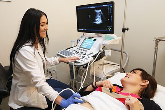
After a brief medical history is taken you will be asked to lie down. Ultrasound examinations are performed by a sonographer. The sonographer will apply a hypoallergenic, water-soluble gel to prevent air from getting between your skin and the transducer. The sonographer then gently passes the transducer over the skin of the area being examined, producing a painless sensation of light pressure. If circumstances dictate, a transvaginal approach may be used. After the exam, the gel is gently wiped off and you may resume normal activities.